 Slit Lamp Anterior Segment Photography is documentation of microscopic and obscure details of the transparent, translucent and opaque structures of the anterior segment and surrounding areas of the eye.
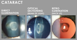
Often conditions affecting the anterior segment of the eye are of a subtle nature and can only be documented using a Slit Lamp Biomicroscope with an attached camera. Slit Lamp Photography utilises a variety of magnifications, angles of view and types of illumination to highlight the areas of interest. This is especially useful in following progression or changes in specific pathology such as new vessels, cataracts and pterygium.
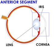
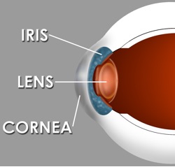 The anterior segment (frontal structures) of the eye include the iris, lens, cornea, eyelids, sclera (white of the eye) and aqueous humor (clear fluid).
Slit Lamp images are used for:
- Comparison
- Pre and Post Surgery Documentation
- Research
- Teaching
- Publication
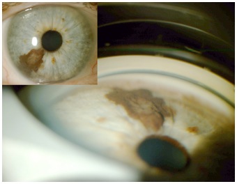
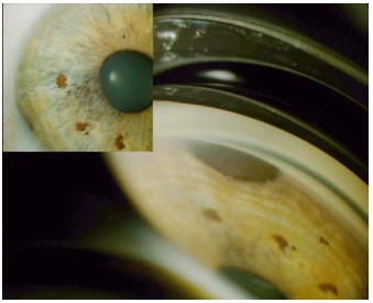
 Gonioscopic Imaging is the use of a goniolens (also known as a gonioscope ) in conjunction with a slit lamp or operating microscope to gain a view of the iridocorneal angle, or the anatomical angle formed between the eye’s cornea and iris.
The gonio lens has an angled mirror that allows images to be taken of structures of the eye that cannot be viewed directly. To perform gonioscopic imaging, eye drops are used to numb the eye.
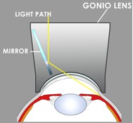 The lens does not rest directly on the cornea, but instead vaults over it, with lubricating fluid filling the gap. The lubricating fluid is used on the lens for comfort, to help maintain a tight lens fit and to prevent the cornea from being scratched.
Slit lamp images are usually taken in conjunction with gonio images to produce a case presentation of the patient’s condition
Goniophotographs are used for:
- Comparison
- Pre and Post Surgery Documentation
- Research
- Teaching
- Publication
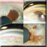
Gonio Image – Inset Slit Lamp Image