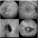
 Indocyanine Green Angiography (ICG) is a diagnostic procedure that uses ICG dye to examine the blood flow in the CHOROID – the layer of blood vessels which lies underneath the retina.
 Indocyanine Green dye is injected into a vein in the arm/hand. As the dye passes through the blood vessels of your eye, photographs are taken to record the blood flow.
The choroidal vessels are hidden beneath a layer of pigmented cells. Infrared light given off by ICG dye can be imaged through the pigmented layer using special filters.
The most common application of indocyanine green angiography is the detection of choroidal neovascularization, a common component of age related macular degeneration.
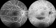
Left: FA Image. Right: ICG Image

What will you experience?
The actual procedure will take 10-20 minutes. The average length of stay in our department can be 1-2 hours.
The Indocyanine Green dye is generally tolerated without any problems. However, ICG dye contains iodine. Severe allergic reactions are possible in people who are allergic to iodine. Our nurse will review your medical history to ensure you are not allergic to substances that contain iodine, such as x-ray dyes and shellfish. ICG dye does not cause urine or skin discoloration.
 Your pupils will be dilated for the ICG.
After the pupils are dilated your vision may become blurred. Driving is not recommended when your pupils are dilated.
Before the procedure
EAT, DRINK & TAKE your medications as you usually do.
DRINK an extra 2-3 cups of water before the procedure.
AVOID coffee, tea, or caffeinated beverages.
BRING an English translator.
BRING your lens case, if you wear contact lenses, as you will need to remove the contacts.