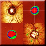
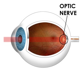
 Heidelberg Retinal Tomography is a diagnostic procedure used for precise observation and documentation of the optic nerve head, essential for the diagnosis and management of glaucoma. The HRT uses a special laser to take 3-dimensional photographs of the optic nerve and surrounding retina. This laser, which will not harm the eye, is focused on the surface of the optic nerve and captures the image.
 The HRT takes images of deeper and deeper layers until the desired depth has been reached. Finally, the instrument takes all these pictures of the layers and puts them together to form a 3D image of the entire optic nerve.
The typical optic nerve damage that occurs in glaucoma is known as “cupping.” As the cells making up the nerve die, due at least in part increased pressure inside the eye, they die and disappear.
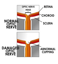 When sufficient numbers of these cells are gone, they leave behind a small “cup” in the nerve. So one important thing doctors look for when they examine the optic nerve is the presence and extent of the “cup,” how deep and wide it is.
The HRT image can be used to compute things such as the area of the optic disc, the volume of the cup, and the area of the rim around the cup. Over several visits, scans are layered and changes are measured.