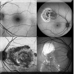
 Autofluorescence Imaging(FAF) is the concept of using naturally occurring fluorescence from the retina to provide an indicator of RPE (layer of the retina) health.
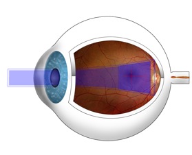
Illuminating the retina with blue light causes certain cellular components to “glow” without injecting any dye. This glow (fluorescence) returning from the retina can be used to create a black-and-white image which can be interpreted by recognizing characteristic patterns, in much the same way clinicians learn to recognize the characteristic patterns in a fluorescein angiogram. ~Heidelberg Engineering
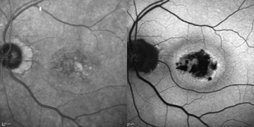
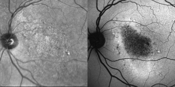
 Fundus autofluorescence patterns may be linked to disease progression in patients with age-related macular degeneration and may thereby help clinicians determine appropriate therapeutic courses in the future.
Potential applications of FAF imaging have been explored in a variety of retinal diseases including: age-related macular degeneration, retinitis pigmentosa, central serous chorioretinopathy and macular dystrophies.
| Standard imaging |
Autofluorescence imaging |
 |
|
 |
|
 |
|