Gregory-Evans Retinal Therapeutics Lab 2011. From left to right: Chris Laver (PhD student); Emran Bashar (PhD student); Kevin Gregory-Evans (PI); Andrew Metcalfe (Research Assistant); Anat Yanai (Senior Research Associate)
The overall theme for the Gregory-Evans Retinal Therapeutics Lab is to identify new ways of treating common retinal diseases, such as macular degeneration and retinitis pigmentosa. This work will focus on novel cell-based therapeutic strategies in model systems (e.g. rodents models of eye disease) and clinical trials. We are undertaking a number of projects to look at how cells can be used to replace damaged retinal tissue and also new ways that cells can be used to deliver and secrete drugs long-term into the diseased retina to prevent further decline of vision.
Past & Present Lab Members
Post-doctoral Research Associates
1. Jim Bellingham (1998-2000)
2. Emma Tartellin (1999-2002)
3. Matt Hodges (1999-2002)
4. Matt Williams (2003-2005)
5. Donna Mackay (2005-2007)
6. Diana Hernandez (2005-2006)
7. Matt Hodges (2006-2008)
8. Anat Yanai (2010-present)
Research Assistants
1. Francis Chang (2007-2009)
2. Kelvin Po (2009-2010)
3. Andrew Metcalf (2010-present)
PhD Students
1. Lindsey Bibb (awarded 2003)
2. Helena Vieira (awarded 2003)
3. James Blackburn (awarded 2004)
4. Julian Patterson (October 2005 – present)
5. AMA Emran Bashar (2011-present)
6. Chris Laver (2011-present)
Collaborators
International:
- John G. Flannery (University of California at Berkeley, USA)
- N.V. Chong (University of Oxford , UK)
- Richard G. Weleber (Oregon Health Sciences University, USA)
National:
- Cheryl Y Gregory-Evans (University of British Columbia)
- Urs O. Hafeli (University of British Columbia)
- Michael Hayden (Simon Fraser University)
- Orson Moritz (University of British Columbia)
- Joanna A. Matsubara (University of British Columbia)
- Marinko V. Sarunic (Simon Fraser University)
- Heather Sheardown (McMaster University)
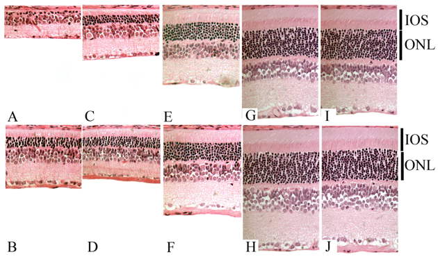
Illustration A. Cell-based drug delivery in the S334ter-4 transgenic model of retinitis pigmentosa. Light microscopy sections at P70. A,B: untreated, C,D : treated with daily subcutaneous gentamicin E,F intravitreal injection of GDNF-secreting mES cells; G,H : combination of both these treatment strategies; I,J : unaffected control Sprague-Dawley rat. Top panels (A,C,E,G,I): sections from upper hemisphere, peripheral retina. Bottom panels (B,D,F,H,J): sections from upper hemisphere, central retina. ONL=outer nuclear layer; IOS=photoreceptor inner and outer segments. ( Ophthal Res 2011;47:32-38).
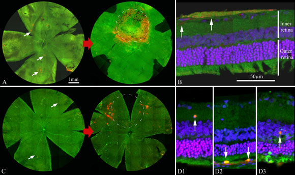
Illustration B. Magnetised stem cells (labeled red) after intravitreal (A,B) or intravenous (C,D) injection, can be localized to a specific retinal locus with a magnet placed within the orbit on the outer surface of the eye (white dashed circle). Cells localize in the inner retina after intravitreal injection (B) but will also accumulate in the outer retina (D) if cells are delivered via intravenous injection. A&C: flatmount rodent retina. B&D: cross-sectional views of treated retina.