Home | About | Background | Research and Projects | Publications | Team | Contact
OCT-A
Segmentation of OCTA using Deep Neural Networks
Accurate segmentation of the retinal microvasculature is a critical step in the quantitative analysis of the retinal circulation, which can be an important marker in evaluating the severity of retinal diseases. Manual segmentation remains the gold standard for segmentation of optical coherence tomography angiography (OCTA) images, but also requires a significant time investment from trained personnel. In our paper Segmentation of the foveal microvasculature using deep learning networks (Prentašic´ et al., 2016) we present a method for automating the segmentation of OCT-A images using deep neural networks (DNNs). Eighty OCT-A images of the foveal region in 12 eyes from 6 healthy volunteers were acquired using a prototype OCT-A system and subsequently manually segmented. The automated segmentation of the blood vessels in the OCT-A images was then performed by classifying each pixel into vessel or non-vessel class using deep convolutional neural networks. When the automated results were compared against the manual segmentation results, a maximum mean accuracy of 0.83 was obtained. When the automated results were compared with inter and intrarater accuracies, the automated results were shown to be comparable to the human raters suggesting that segmentation using DNNs is comparable to a second manual rater. As manually segmenting the retinal microvasculature is a tedious task, having a reliable automated output such as automated segmentation by DNNs, is an important step in creating an automated output.
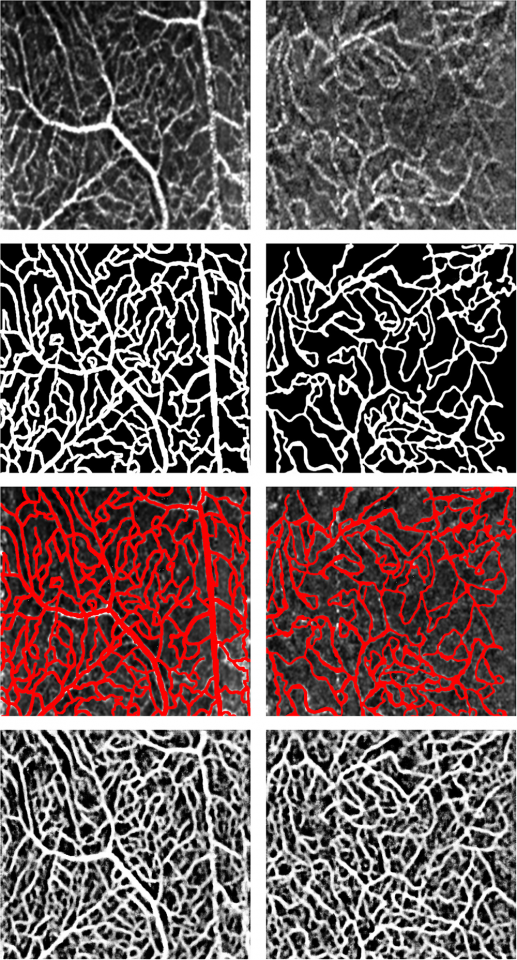
Figure: Examples of OCT-A retinal images acquired with our system
(top row), manual segmentations of the vessels (second row), original
images with manual segmentations superimposed (third row), and
outputs of the proposed DNN method (bottom row). Images in the
left column represent an example of a typical dataset and images
in the right column represent an example of a low quality dataset.
Sequential Image Motion Corrected Averaging
The visibility of retinal microvasculature in optical coherence tomography angiography (OCT-A) images is negatively affected by the small dimension of the capillaries, pulsatile blood flow, and motion artifacts. Serial acquisition and time-averaging of multiple OCT-A images can enhance the definition of the capillaries and result in repeatable and consistent visualization. In our paper Strip-based registration of serially acquired optical coherence tomography angiography (Heisler et al., 2017) we demonstrate an automated method for registration and averaging of serially acquired OCT-A images. Ten OCT-A volumes from six normal control subjects were acquired using our prototype 1060-nm swept source OCT system. The volumes were divided into microsaccade-free en face angiogram strips, which were registered to a template image. The resulting averaged images were presented of all the retinal layers combined, as well as in the superficial and deep plexus layers separately. The improved visualization of the capillaries can enable robust quantification and study of minute changes in retinal microvasculature.
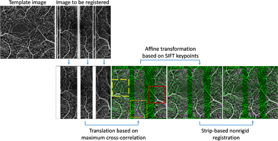
Figure: Demonstration of the image stripping, coarse translation, affine registration, and nonrigid registration steps of the proposed algorithm. The template image (green) and registered strip (magenta) are shown as composite images where white regions indicate where the two images have the same intensities.
http://biomedicaloptics.spiedigitallibrary.org/article.aspx?articleid=2610232
Imaging the Foveal Avascular Zone in Retinopathy of Prematurity (ROP) Patients
Spectral domain OCT (SD-OCT) is capable of showing microstructural abnormalities in retinas of patients born prematurely. OCT Angiography (OCTA), on the other hand, can show vascular changes in common retinal and choroidal disease. Thus, it is possible that patients with retinopathy of prematurity (ROP), a disease that causes abnormal blood vessels to grow in the retina, could have irregularities in their foveal architecture that are not recognized on SD-OCT. In our study Optical Coherence Tomography Angiography in Retinopathy of Prematurity versus Normal Eyes (Teja et al., 2017), we evaluated foveal avascular zone (FAZ) parameters and perifoveal capillary density (PCD) of OCTA images in ROP eyes and normal control eyes, with the motivation that OCTA would be able to visualize abnormalities that are not otherwise recognized on clinical examination or with SD-OCT/FA. Our findings show that OCTA is advantageous in imaging the superficial and deep capillary plexuses, and that ROP patients have a smaller FAZ. However, there were challenges in acquiring images of ROP patients, which led to images that were partially degraded by motion artifact.
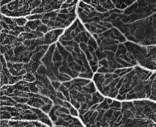
Figure: OCTA image of an ROP patient. The foveal avascular zone is significantly diminished.
Sensorless AO-OCT
Optical coherence tomography (OCT) has revolutionized modern ophthalmology, providing depth resolved images of the retinal layers in a system that is suited to a clinical environment. Although the axial resolution of an OCT system is sufficient to resolve retinal features at a micrometer scale, the lateral resolution is dependent on the delivery optics and is limited by ocular aberrations. This has been our motivation for developing the wavefront-sensorless adaptive optics system currently in our laboratory. The resolution afforded by the WSAO system will allow us to visualize the photoreceptor mosaic as well as individual photoreceptors. We will be testing the system in a clinical setting in the near future, and are eager to share updates on our progress. Below are two images from a previous study we performed with a WSAO system, Lens-based wavefront sensorless adaptive optics swept source OCT (Jian et al., 2016).
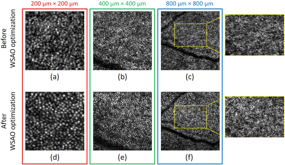
Photoreceptor images before (a–c) and after (d–f) WSAO aberration correction acquired at three different fields of view: (a) and (d), 200 μm × 200 μm; (b) and (e), 400 μm × 400 μm; and (c) and (f), 800 μm × 800 μm.
Y. Jian, S. Lee, M.J. Ju, M. Heisler, W. Ding, R.J. Zawadzki, S. Bonora, M.V. Sarunic, “Lens-based wavefront sensorless adaptive optics swept source OCT,” Scientific Reports 6, (2016): 27620.
https://www.nature.com/articles/srep27620
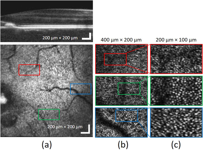
Figure 2: Photoreceptor cone mosaic images acquired at different retinal eccentricities (2.3°, 3.3° and 5.7° ); (a) wide-field OCT B-scan and en face images, (b) photoreceptor images with 400 μm × 200 μm field of view, (c) photoreceptor images with 200 μm × 100 μm field of view.
Y. Jian, S. Lee, M.J. Ju, M. Heisler, W. Ding, R.J. Zawadzki, S. Bonora, M.V. Sarunic, “Lens-based wavefront sensorless adaptive optics swept source OCT,” Scientific Reports 6, (2016): 27620.
https://www.nature.com/articles/srep27620